K-State Technology Development Institute partners with veterinary college to develop 3D-printed animal eyes for ophthalmology training
Thursday, March 7, 2024

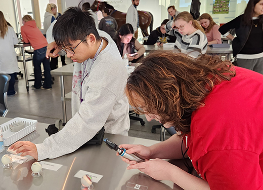
K-State's Technology Development Institute and College of Veterinary Medicine teamed up to create 3D-printed animal eye models for veterinary students to use when training for fundoscopy exams. | Download this photo.
MANHATTAN — Kansas State University veterinary students have a new and improved way to study veterinary ophthalmology thanks to a collaboration between the College of Veterinary Medicine and the Technology Development Institute.
The Technology Development Institute, or TDI, and the College of Veterinary Medicine developed a new training aid to help veterinary students learn to use eye exam equipment on several species of animal patients: 3D-printed eye globes replicating the eyes of dogs, cats, horses and rabbits.
These model eye globes allow the students to practice using real equipment to perform direct and indirect fundoscopy exams to check the fundus — the back of the inside of the eye, including the retina and optic nerve.
The models are based on photographic images captured by the ophthalmologists at the College of Veterinary Medicine. The engineering team at TDI designed a two-part round assembly with a clear lens and cornea on one half of the globe and the fundus image 3D printed on the inside of the other half of the globe. The two halves are combined to form a model eye training aid that enables students to learn the coordination skills of an ophthalmoscope, the instrument used for inspecting the eye.
The idea for the training aid was developed by K-State's Susan Rose, clinical skills instructor, and Shane Lyon, clinical associate professor and clinical skills coordinator, both in the College of Veterinary Medicine. Rose and Lyon teach freshman and sophomore veterinary students in courses that introduce the hand skills required to perform various tasks in a low-stakes setting.
One goal for these courses has been to reduce the use of live animals in teaching these base skills. Using models like the eye globes, students can learn the coordination and hand skills required and develop muscle memory with a realistic replica that doesn't require a live animal. Once the students' foundational skills are established, they have the knowledge they need when met with the added challenge of a live patient.
Previous attempts at creating a model eye for the lab fell short of meeting the instructors' needs. When Rose learned of TDI's ability to 3D print photographic images in high resolution, it immediately led to the idea of creating this model set, which would dramatically improve the ability to teach these skills.
"What we needed was a model that provided not just representative images of a normal fundus but one that could teach them how to utilize more functions of the ophthalmoscope when doing direct fundoscopy," Rose said. "I wanted to have them learn how to use the diopter to focus on different planes, not just look at the back of the eye. This was a significant improvement over their previous handmade models."
Rose said TDI's model provides a better learning opportunity for students because it includes the mid-range iris and the clear lens cornea — the outer layer of the eye — in addition to the fundus, unlike previous models.
The initial training eyes are extra large to make it easier for students to learn the more challenging indirect fundoscopy. This technique requires them to aim a light source through the dilated pupil, then drop down a special hand-held magnifying lens to view the fundus.
Quinton Berggren, senior engineer at TDI, worked closely with Rose and Lyon on the design of the eye globe to ensure it was an accurate representation of the animal and would meet the needs of the vet students. Jessica Meekins, associate professor of ophthalmology, and Amy Rankin, professor of ophthalmology, also provided recommendations to refine the model, including advice about the specific animal globes that should be included in the set.
"Working with the College of Veterinary Medicine enables us to do things with our 3D printers that we would have never imagined were possible," Berggren said. "They have been able to provide us with all of the details that we need to be able to design and produce the materials as required, and that is what makes this a fun and unique partnership."
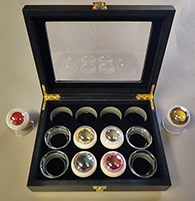
To date, the team has developed two canine, two feline, one equine and one lagomorph —rabbit — eye globes, and the globes have been packaged into a single training kit product. These training kits are being offered for sale to other veterinary schools, and the team is working to identify distribution partners who can help market and promote the training aids globally.
This project was completed in support of the K-State 105 initiative, Kansas State University's answer to the call for comprehensive economic growth and advancement solutions for Kansas. Learn more at k-state.edu/105.
The K-State Technology Development Institute in the Carl R. Ice College of Engineering is a U.S. Department of Commerce Economic Development Administration University Center and received a grant from the Research and Entrepreneurship Federal Matching Grant Dollars Fund. TDI provides a broad range of engineering and business development services to both private industry and university researchers to advance the commercial readiness of new products or technologies.

